The dura mater is composed of two layers: the periosteal/endosteal layer and the meningeal layer. The dural venous sinuses are between these two layers. The dura folds to form septa that create the falx cerebri
What layer is the dura mater?
Dura mater is the outermost layer of the meninges. It has its vascular supply. The dura mater’s meningeal layer bends towards the inner direction of itself to form 4 structural forms, called the dural reflections.
Which layer is the dura mater quizlet?
1) Dura mater – the dura mater layer is the thickest, outermost layer.
What are the three layers of the dura mater?
Dura materFMA9592Anatomical terminologyWhat is formed when the two layers of dura separate?
The two dural layers separate at certain sites to form venous sinuses. In addition, reflections of the inner meningeal layer form septa that divide the brain into compartments (Fig. 2). The largest reflection, the falx cerebri, is formed by midline reflections from the top of the calvarium.
What are the 3 layers of the brain?
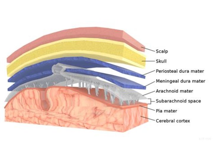
Three layers of membranes known as meninges protect the brain and spinal cord. The delicate inner layer is the pia mater. The middle layer is the arachnoid, a web-like structure filled with fluid that cushions the brain. The tough outer layer is called the dura mater.
Is dura mater avascular?
Epidural Space and Dura Mater While it was once thought that the dura was avascular, it is actually highly vascular in nature, as the major vessels that supply it run in the epidural space deep to the skull.
What is underneath the arachnoid layer?
The space under the arachnoid, the subarachnoid space, is filled with cerebrospinal fluid and contains blood vessels. The pia mater is the innermost layer of meninges. This thin, delicate membrane is tightly bound to the surface of the brain and spinal cord and cannot be dissected away without damaging the surface.What are the dural folds?
The meningeal layer of the dura mater creates several dural folds that divide the cranial cavity into freely communicating spaces. The function of the dural folds is to limit the rotational displacement of the brain. The folds include the following: The falx cerebri is a meningeal projection of dura in the brain.
What is the outer layer of the brain called?Brain Coverings: Meninges Three layers of protective covering called meninges surround the brain and the spinal cord. The outermost layer, the dura mater, is thick and tough. It includes two layers: The periosteal layer of the dura mater lines the inner dome of the skull (cranium) and the meningeal layer is below that.
Article first time published onWhere is the dura mater?
The tough outer layer of tissue that covers and protects the brain and spinal cord and is closest to the skull. The dura mater is one of the three layers that form the meninges.
What is dura mater quizlet?
dura mater. Thick, outermost layer of the meninges surrounding and protecting the brain and spinal cord.
What is DURA?
Dura: The outermost, toughest, and most fibrous of the three membranes (meninges) covering the brain and the spinal cord. Dura is short for dura mater (from the Latin for hard mother). … An accumulation of blood outside the dura is an epidural hematoma. Subdural means under the dura.
What is the difference between the periosteal and meningeal layers of the dura mater?
Dura mater that surrounds the brain consists of two layers. The outer layer is called the periosteal layer and the inner layer is the meningeal layer. The outer periosteal layer firmly connects the dura mater to the skull and covers the meningeal layer. The meningeal layer is considered the actual dura mater.
What is the difference between dura mater and arachnoid mater?
The dura mater is a thick, tough, and durable membrane composed of dense fibrous connective tissue. … The arachnoid mater is a very thin and transparent membrane that lies on top of a fluid filled space directly inferior to the membrane. This space, the subarachnoid space, is filled with cerebropinal fluid.
In what way is the dura mater of the brain different from the dura mater of the spinal cord quizlet?
In what way is the dura mater of the brain different from the dura mater of the spinal cord? The dura mater of the brain is made of two separate layers. Which of the following is NOT a function of the specialized ependymal cells that form the choroid plexus?
How many layers does the brain have?
There are 3 layers of tissue called meninges that help protect the brain. The outer covering of tissue (called the dura mater), closely lines the inside of the skull. The second layer is the arachnoid mater, and the third layer, the pia mater, hugs the surface of the brain.
Does subarachnoid space lies between what two layers of meninges?
Anatomically, the subarachnoid space exists between the arachnoid mater externally and pia mater internally. A network of fine delicate connective tissue called trabeculae connects these two layers and gives this space its characteristic spider web appearance.
What are the cranial dural septa?
–Extensions of meningeal dura mater into the cranial cavity. …
What is the Dura in the brain?
The dura mater often gets referred to as merely the dura. It is one of the layers of connective tissue that make up the meninges of the brain (pia, arachnoid, and dura, from inside to outside). It is the outermost layer of the three meninges that surround and protect the brain and spinal cord.
What are the major layer of the brain?
The brain is split up into three major layers: the hindbrain, the midbrain, and the forebrain.
What is the dura mater what is its function?
The dura mater is a sac that envelops the arachnoid and has been modified to serve several functions. The dura mater surrounds and supports the large venous channels (dural sinuses) carrying blood from the brain toward the heart. The dura mater is partitioned into several septa, which support the brain.
How many dural folds are there?
The two layers are separated by the dural venous sinuses. From these, the meningeal layer extends into the cranial cavity and forms four main dural folds, also known as dural reflections (Rea, 2015):
What is the pia mater?
The pia mater is the meningeal envelope that firmly adheres to the surface of the brain and spinal cord. It is a very thin membrane composed of fibrous tissue covered on its outer surface by a sheet of flat cells thought to be impermeable to fluid.…
What was attached to the dura mater?
The dura mater attaches to the periosteum and accompanies the optic nerve as far as the orbit. It forms a recess, which is concave posteriorly, called the tent of the optic nerve, running from the sphenoidal limbus to the posterior clinoid process.
What is spiral cord?
A column of nerve tissue that runs from the base of the skull down the center of the back. It is covered by three thin layers of protective tissue called membranes. The spinal cord and membranes are surrounded by the vertebrae (back bones).
Which place is located between arachnoid mater and pia mater?
The subarachnoid space is the space that normally exists between the arachnoid and the pia mater. It is filled with cerebrospinal fluid and continues down the spinal cord.
What are the folds of the brain called?
As shown in figure 1a (bottom), the human brain exhibits an intricate pattern of convex folds (gyri) and valleys (sulci). The first, or primary, folds emerge in consistent locations across individuals and between species.
What are the two hemispheres of the brain?
The cerebrum is divided into two major parts: the right and left cerebral hemispheres or halves at a fissure, the deep groove down the middle. The hemispheres communicate with each other through the corpus callosum which is a bundle of fibers between the hemispheres.
What are the folds and valleys that make the brain look wrinkled?
The “valleys” of the wrinkles are called sulci (or sometimes, fissures); the “peaks” between wrinkles are called gyri.
What are the two main functions of the CSF?
CSF provides hydromechanical protection of the neuroaxis through two mechanisms. First, CSF acts as a shock absorber, cushioning the brain against the skull. Second, CSF allows the brain and spinal cord to become buoyant, reducing the effective weight of the brain from its normal 1,500 grams to a much lesser 50 grams.