The gallbladder mucosa is lined with a single layer of columnar epithelial cells that, in many species, secondarily form prominent folds that can reach into the muscular layer, forming Rokitansky–Aschoff sinuses.
What type of cells are in the gallbladder?
The inner surface of the gall bladder is covered by the mucosa. The sufrace is made up of a simple columnar epithelium. The epithelial cells have microvilli, and look like absorptive cells in the intestine. Underneath the epithelium is the lamina propria.
What do gallbladder cells produce?
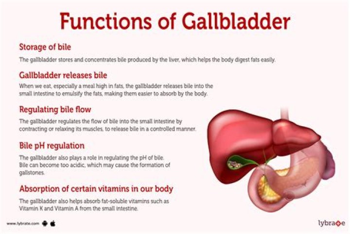
However, not all bile runs directly into the duodenum. About 50% of the bile produced by the liver is first stored in the gallbladder. This is a pear-shaped organ located directly below the liver. Then, when food is eaten, the gallbladder contracts and releases stored bile into the duodenum to help break down the fats.
What is the lining epithelium of bile duct?
The intrahepatic ducts, cystic duct, and the common bile duct are lined by a tall columnar epithelium. The gallbladder stores bile excreted from the liver. The columnar mucosa is arranged in folds over the lamina propria, allowing expansion.What color is the mucosa in gallbladder?
Gross photograph of gallbladder mucosa, which is soft and velvety (as well as bright green when fresh).
Where is simple squamous epithelium found?
Simple squamous epithelium Simple squamous epithelia consist of a single layer of flattened cells. This type of epithelia lines the inner surface of all blood vessels (endothelium), forms the wall of alveolar sacs in the lung and lines the body cavities (mesothelium).
What is a Cholangiocyte?
Cholangiocytes are a heterogeneous, highly dynamic population of epithelial cells that line a three-dimensional network of bile ducts known as the biliary tree. Their major physiologic function lies in modification of hepatic canalicular (i.e., primary) bile as it is transported along the biliary tree.
What is the columnar epithelium?
The columnar epithelium is composed of epithelial cells that are column-shaped. The cell comprising the columnar epithelium is taller than it is wide. Its height is approximately four times its width. The nucleus in each cell is elongated and often found near the base.What is a stratified squamous epithelium?
Stratified squamous epithelia have two or more layers of cells, with a superficial squamous layer and basal layers that are usually cuboidal or columnar. … This type of epithelium can withstand abrasion because the loss of cells from the surface does not compromise the underlying tissue.
What is the bile produced?Bile is a fluid that is made and released by the liver and stored in the gallbladder. Bile helps with digestion. It breaks down fats into fatty acids, which can be taken into the body by the digestive tract.
Article first time published onHow does your gallbladder produce bile?
The gallbladder acts as a storage vessel for bile produced by the liver. Bile is produced by hepatocytes cells in the liver and passes through the bile ducts to the cystic duct. From the cystic duct, bile is pushed into the gallbladder by peristalsis (muscle contractions that occur in orderly waves).
What is bile made up of?
Bile consists of ~95% water in which are dissolved a number of endogenous solid constituents including bile salts, bilirubin phospholipid, cholesterol, amino acids, steroids, enzymes, porphyrins, vitamins, and heavy metals, as well as exogenous drugs, xenobiotics and environmental toxins (76).
What is the mucus membrane?
Listen to pronunciation. (MYOO-kus MEM-brayn) The moist, inner lining of some organs and body cavities (such as the nose, mouth, lungs, and stomach). Glands in the mucous membrane make mucus (a thick, slippery fluid).
What Innervates the gallbladder?
The gallbladder receives parasympathetic nerve supply from the right vagus through its hepatic branch; sympathetic supply comes from T 7-9 through the celiac plexus.
Why submucosa is absent in gall bladder?
SUBMUCOSA OF GALL BLADDER A MISCONCEPTION Muscularis mucosa is totally absent in the wall of the gall bladder. The lamina propria containing loose connective tissue rest upon the muscularis externa and hence there is no muscular layer separating the mucosa from the muscularis externa.
What is a stellate cell?
The stellate cell, previously known as the Ito cell, fat-storing cell, perisinusoidal cell or lipocyte, is a major storage site for vitamin A. In liver injury, it becomes a transitional cell or myofibroblast-like cell capable of synthesising collagen types I, III and IV as well as laminin.
What is Alagille syndrome?
Alagille syndrome is an inherited condition in which bile builds up in the liver because there are too few bile ducts to drain the bile. This results in liver damage. Your liver makes bile to help remove waste from your body. It also helps digest fats and the fat-soluble vitamins A, D, E, and K.
What is the hepatic triad?
por·tal tri·ad. (pōr’tăl trī’ad) Branches of the portal vein, hepatic artery, and the biliary ducts bound together in the perivascular fibrous capsule or portal tract as they ramify within the substance of the liver.
What are the 4 types of epithelial tissue?
In general, simple epithelial tissues are classified by the shape of their cells. The four major classes of simple epithelium are (1) simple squamous, (2) simple cuboidal, (3) simple columnar, and (4) pseudostratified.
Which one of the following cell types is found in epithelial tissue?
There are three principal cell shapes associated with epithelial cells: squamous epithelium, cuboidal epithelium, and columnar epithelium.
What are squamous epithelial tissues?
Squamous means scale-like. simple squamous epithelium is a single layer of flat scale-shaped cells. Both the endothelial lining of blood vessels and the mesothelial lining of the body cavities are simple squamous epithelium.
Does mucosal stratified squamous epithelium have a stratum corneum?
It consists of two layers, the surface stratified squamous epithelium and the deeper lamina propria. In keratinized oral mucosa, the epithelium is composed of the four layers stratum basale, stratum spinosum, stratum granulosum, and stratum corneum.
Is keratin found in stratified squamous and not in other epithelial tissue types?
The cells on the surface of stratified squamous keratinized epithelium are very flat. Not only are they flat, but they are no longer alive. They have no nucleus or organelles. They are filled with a protein called keratin, which is what makes our skin waterproof.
What is the Keratinized epithelium?
Keratinized epithelium, is composed of numerous layers of dead squamous cells, which are specially structured to be waterproof and reduce evaporation from underlying tissues. Therefore they constitute an important part of the epidermis or external skin.
What cells make up simple columnar epithelium?
Simple Columnar Epithelium is made up of Glandular Goblet cells which secrete mucins to form mucin. the rest of the cell is made up of cytoplasm with membrane bound secretory granules which secrete the mucin, and are found towards the apical surface of the cell.
What are columnar epithelial and goblet cells?
Simple columnar epithelial cells absorb material from the digestive tract. Goblet cells secret mucous into the digestive tract lumen. Columnar epithelial cells are taller than they are wide: they resemble a stack of columns in an epithelial layer, and are most commonly found in a single-layer arrangement.
How many types of epithelial tissue are there?
There are 8 types of epithelial tissues. Simple squamous, Stratified Squamous, Simple Cuboidal, Stratified Cuboidal, Simple Columnar, Stratified Columnar, Pseudostratified Columnar and Transitional epithelia or urothelium.
What is villi what is their location and function?
What is their location and function? Solution 5: Villi are small finger-like projections found inside the inner walls of the small intestine. They v increase the surface area for absorption of the digested food. Each villus has a network of thin and small blood vessels close to its surface.
Is bile a poop?
Stool color is generally influenced by what you eat as well as by the amount of bile — a yellow-green fluid that digests fats — in your stool. As bile pigments travel through your gastrointestinal tract, they are chemically altered by enzymes, changing the pigments from green to brown.
Does liver produce bile?
Your liver continually produces bile. This is a chemical that helps turn fats into energy that your body uses. Bile is necessary for the digestive process.
Where is the sphincter of Oddi?
The sphincter of Oddi refers to the smooth muscle that surrounds the end portion of the common bile duct and pancreatic duct. This muscle relaxes during a meal to allow bile and pancreatic juice to flow into the intestine.