The space beneath the flexor retinaculum is divided into four separate tunnels. Three tendons, and the posterior tibial vessels and nerve, pass through these tunnels as they pass around the ankle and into the foot.
What passes through the flexor retinaculum of the foot?
The flexor retinaculum of the foot extends from the medial malleolus above, to the calcaneus below. This converts a series of bony grooves into canals for the passage of the tendons of the flexor muscles and the posterior tibial vessels and tibial nerve into the sole of the foot, known as the tarsal tunnel.
What muscles attach to flexor retinaculum?
The tendons of the palmaris longus and flexor carpi ulnaris are partly attached to the surface of the retinaculum; below, the short muscles of the thumb and little finger originate from the flexor retinaculum.
What passes through peroneal Retinaculum?
The peroneal retinaculum is divided into the superior and inferior peroneal retinaculum 1. They house the tendons of fibularis longus and brevis as they pass through the lateral aspect of the ankle joint posterior to the lateral malleolus 1.What is the function of flexor retinaculum?
The flexor retinaculum forms a retinacular bridge over the carpal tunnel extending from ulnar to radial direction. Its main function is to protect the contained without a significant mechanical action in supporting the transverse carpal arch.
Is the peroneal tendon a flexor or extensor?
Answer-peroneals are considered “flexors” or evertors, the AMA recently confimed to her, after consulting with a CPT advisor from the American Orthopaedic Foot and Ankle Society. Of the peroneal tendons, only the peroneus tertius tendon has “extensor” capability.
What is the main purpose of the retinaculum at the ankle?
A retinaculum is a band of thick deep fascia that holds the long tendons of your ankle (those that cross the ankle) in place. Retinaculum also acts as a pulley system increasing mechanical advantage. Retinaculum are a major source of neurological receptors involved in balance and proprioception.
Is the superior peroneal Retinaculum a ligament?
The fibers of the superior retinaculum (external annular ligament) are attached above to the lateral malleolus and below to the lateral surface of the calcaneus.What is the function of the peroneal tendon?
The main function of the peroneal tendons is to stabilize the foot and ankle and protect them from sprains.
Which structure passes superficial to flexor retinaculum?Structures passing superficial to flexor retinaculum of the hand are: Ulnar nerve. Ulnar artery and vein. Palmar cutaneous branch of ulnar nerve.
Article first time published onWhich bone is attached to flexor and extensor Retinaculum?
These four bony points are all palpable in the living hand and it should be noted that pisiform is the only carpal bone that gives attachments to both flexor and extensor retinacula.
What happens when you cut the flexor retinaculum?
When the FR is cut in longitudinal direction over the median nerve, the median nerve elevates between the two borders of the retinaculum and the scar tissue between the median nerve and the flexor retinaculum leads to nerve constriction.
What passes through carpal tunnel?
A passageway from the wrist to the hand, the carpal tunnel is made of tendons, ligaments and bones. The median nerve passes through the tunnel and provides sensation to your thumb, index finger, middle finger and the thumb side of the ring finger.
What is a Retinaculum tear?
In type II injuries, the superior peroneal retinaculum is torn off its attachment to the fibula. In type III injuries, a small bony fragment avulses off the distal fibula along with the superior peroneal retinaculum. In type IV injuries, the retinaculum is torn off its posterior attachment to the calcaneus.
What passes under Sustentaculum Tali?
At the upper and forepart of the medial surface of the calcaneus, below the middle talar facet, there is a horizontal eminence, the talar shelf (also sustentaculum tali), which gives attachment to the plantar calcaneonavicular (spring) ligament, tibiocalcaneal ligament, and medial talocalcaneal ligament.
Can you tear your flexor retinaculum?
Medial flexor retinaculum injuries are not uncommon, but medial flexor retinaculum periosteal avulsion injuries are rare. This patient sustained a medial flexor retinaculum tear readily characterized at computed tomography by an associated proximal retinacular avulsion fracture from the posteromedial tibia.
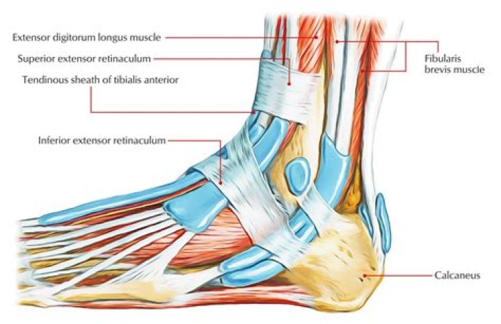
What is the function of the superior extensor Retinaculum?
The structure indicated is the superior extensor retinaculum of the leg. Retinacula (retinaculum singular) are bands of connective tissue which surround tendons and hold them in place. They function to stabilise tendons as the muscles to which they attach contract to cause movement.
What is superior peroneal Retinaculum?
The superior peroneal retinaculum (SPR) functions as the primary restraint to peroneal tendon subluxation and is also a secondary restraint to anterolateral ankle instability. It is formed from a confluence of the common peroneal sheath and the superficial fascia of the leg.
Where do peroneal tendons insert?
The superior peroneal retinaculum covers the peroneal muscle tendons with attachments at the distal fibula and lateral calcaneus. The inferior peroneal retinaculum covers the peroneal muscle tendons with insertions at the lateral calcaneus (interior to the superior peroneal retinaculum).
What is a dislocated peroneal tendon?
Tough bands of tissue (peroneal retinaculum) pass over the peroneal tendons to keep them in this position. If these bands are damaged, the peroneal tendons are able to move out of their groove. This is called a dislocation of the peroneal tendons.
What is the tendon on the outside of your ankle?
The peroneal tendons are on the outside of the ankle just behind the bone called the fibula. Peroneal tendinosis is the name for the enlargement, thickening, and swelling of these tendons.
How do you know if you have peroneal tendonitis?
- pain at the back of the ankle.
- pain that worsens during activity and lessens during rest.
- pain when turning the foot in or out.
- swelling at the back of the ankle.
- instability of the ankle when bearing weight.
- the area is warm to the touch.
Should you massage peroneal tendonitis?
Massage. Your therapist may use soft tissue massage techniques to improve peroneal tendon mobility on the lateral side of your ankle. Massage may help improve tissue flexibility and circulation, and it may be used prior to exercise and stretching to improve overall mobility.
What causes peroneal tendonitis?
What causes peroneal tendonitis? Peroneal tendon inflammation can develop over time with repetitive overuse of the tendons. Or it might happen suddenly due to an acute ankle injury like a sprain. The tendons or the lubricated sheath that surrounds the tendons can swell, making it hard for them to move smoothly.
Which two structures pass through the tunnel of Guyon?
The ulnar nerve, together with the ulnar artery, passes through the tunnel of Guyon. This tunnel lies between two dynamic structures, the pisiform and hamate bones, and is covered by the pisohamate ligament (Fig. 26), which is a continuation of the flexor carpi ulnaris tendon.
Where is flexor digitorum Superficialis?
Flexor Digitorum Superficialis along with other superficial muscles of volar compartment of forearm. It is the bulk of muscle located at the superficial volar/anterior aspect of the forearm.
Which structures pass posterior to the flexor retinaculum?
- Median nerve.
- Radial bursa.
- Ulnar bursa.
What goes under the extensor retinaculum of hand?
Deep to extensor retinaculum, its tendons are placed between the tendons of extensor digiti minimi on its medial side, and extensor pollicis longus on its lateral side. … In the dorsum of the hand the tendons of extensor digitorum run superficial to dorsal interossei muscles.
Where does extensor retinaculum attach?
The extensor retinaculum is attached laterally to the lateral margin of the radius. However, it is not attached to the ulna, as the distance between these two bones varies with supination and pronation of the forearm.
What travels through the tendon of the flexor digitorum profundus?
It is one of two flexor muscles that is not exclusively supplied by the median nerve (the other is flexor carpi ulnaris). In the forearm, the median nerve travels distally between the flexor digitorum superficialias and the flexor digitorum profundus.
What goes through Guyon's canal?
Guyon’s canal also called ulnar tunnel or ulnar canal, is an anatomical fibro-osseous canal located on the medial side of the hand. … The ulnar nerve and ulnar artery pass through the Guyon canal as they pass from distal forearm to the hand.