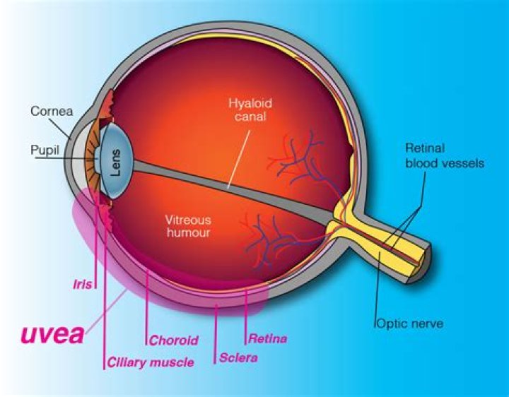
The uvea consists of the layer and structures of the eye beneath the white of the eye
Is the choroid the uvea?
The uvea or vascular tunic of the eye consists of the choroid, ciliary body, and iris. The choroid lies between the sclera and RPE, and contains connective tissue, capillaries, and melanocytes. The choroid terminates anteriorly as the ciliary body.
Where is uvea attached to sclera?
It has a rough outer surface which is attached to the sclera at the optic nerve and at the exit of the vortex veins. The smooth inner surface of the choroid is attached to the retinal pigmented epithelium (RPE). Choroid becomes continuous with pia and arachnoid at the optic nerve.
What does the uvea include?
The uvea is the middle layer of the eye. It lies beneath the white part of the eye (the sclera). It is made of the iris, ciliary body, and choroid. These structures control many eye functions, including adjusting to different levels of light or distances of objects.What is difference between choroid and uvea?
is that choroid is (anatomy) the vascular layer of the eye lying between the retina and the sclera while uvea is (anatomy) the middle of the three concentric layers that make up the eye; it is pigmented and vascular, and comprises the choroid, the ciliary body, and the iris.
What is the uvea of the eye?
The uvea is the middle layer of tissue in the wall of the eye. It consists of the iris, the ciliary body and the choroid. When you look at your eye in the mirror, you will see the white part of the eye (sclera) and the colored part of the eye (iris). The iris is located inside the front of the eye.
Is Pars Plicata part of uvea?
It is divided into two parts: a thicker body (pars plicata) anterior to a flatter body (pars plana). The pars plana of the ciliary body is then continuous with the most posterior component of uvea, the choroid. The choroid extends from termination of the pars plana at the ora serrata to the optic nerve.
Where is ciliary muscle?
The ciliary muscle is elongated, triangular in shape, and located beneath the anterior sclera just posterior to the limbus. The shortest side of the triangular region faces anterior-inward and it is to this region of the ciliary body that the base of the iris inserts.Is the uvea blue?
The middle coat of the eye is called the uvea (from the Latin for “grape”) because the eye looks like a reddish-blue grape when the outer coat has been dissected away.
Why is it called uveal tract?The uveal tract, or simply uvea, is the pigmented middle membrane of the layers that make up the eye. The uveal tract is also called the vascular tunic of the eye because it is rich in its blood supply – i.e., vascular – and because it envelops the eye like a tunic would cover a body.
Article first time published onWhat does uvea mean in Latin?
FMA. 58103. Anatomical terminology. The uvea (/ˈjuːviə/; Lat. uva, “grape”), also called the uveal layer, uveal coat, uveal tract, vascular tunic or vascular layer is the pigmented middle of the three concentric layers that make up an eye.
What is the posterior uvea?
Definition. Posterior uveitis is inflammation of the back part of the uvea known as the choroid. The uvea is the middle layer of the eye. Early treatment can improve outcomes. Normal Anatomy of the Eye.
Which part of the eye is responsible for focusing light?
The iris (the colored part of the eye) controls how much light the pupil lets in. Next, light passes through the lens (a clear inner part of the eye). The lens works together with the cornea to focus light correctly on the retina.
What types of pathological processes of uvea do you know?
Common pathologic changes involving the uvea include inflammatory and neoplastic diseases. Inflammatory changes are clinically recognized as various forms of uveitis. Among the neoplasms, both primary and metastatic tumors are found in all parts of the uvea.
Which area has no vision or sight?
blind spot, small portion of the visual field of each eye that corresponds to the position of the optic disk (also known as the optic nerve head) within the retina. There are no photoreceptors (i.e., rods or cones) in the optic disk, and, therefore, there is no image detection in this area.
Is ciliary body transparent?
The ciliary body is a ring-shaped thickening of tissue inside the eye that divides the posterior chamber from the vitreous body. … The inner layer is transparent and covers the vitreous body, and is continuous from the neural tissue of the retina.
Where is the Pars Plicata?
The pars plicata is located anterior to the pars plana portion of the ciliary body, and posterior to the iris.
What is pupil?
Listen to pronunciation. (PYOO-pul) The round opening in the center of the iris (the colored tissue that makes the “eye color” at the front of the eye). The pupil changes size to let light into the eye.
Where is the aqueous Humour produced?
Aqueous humor is produced by the ciliary body, in uveoscleral route, it flows from the posterior chamber through the pupil into the anterior chamber and then (shown by dashed lines and arrowheads) through the face of the ciliary body and iris root to the ciliary muscle and suprachoroidal space to either veins in the …
How long does it take to go blind from uveitis?
The mean duration of visual loss was 21 months. Of the 148 patients with pan-uveitis, 125 (84.45%) had reduced vision, with 66 (53%) having vision ⩽6/60.
Is iritis the same as uveitis?
Iritis (i-RYE-tis) is swelling and irritation (inflammation) in the colored ring around your eye’s pupil (iris). Another name for iritis is anterior uveitis. The uvea is the middle layer of the eye between the retina and the white part of the eye.
Does uveitis ever go away?
Some forms of uveitis take a long time to go away. Some come back after treatment. Depending on the disease type, treatments include: Antibiotics, antivirals or antifungals: These medications treat uveitis caused by an infection.
What is the white of the eyes?
The sclera is the dense connective tissue of the eyeball that forms the “white” of the eye. It is continuous with the stroma layer of the cornea. The junction between the white sclera and the clear cornea is called the limbus.
What is a ciliary flush?
Ciliary flush is usually present in eyes with corneal inflammation, iridocyclitis or acute glaucoma, though not simple conjunctivitis. A ciliary flush is a ring of red or violet spreading out from around the cornea of the eye.
What are the ciliary muscle?
Ciliary muscle: A circular muscle that relaxes or tightens the zonules to enable the lens to change shape for focusing. The zonules are fibers that hold the lens suspended in position and enable it to change shape during accommodation.
What is the ciliary ganglion?
Ciliary ganglion is a peripheral parasympathetic ganglion. It is situated near the apex of orbit between the optic nerve and lateral rectus muscle. It is related medially to the ophthalmic artery and laterally to the lateral rectus muscle.
What is the main function of the ciliary muscles?
The ciliary body produces the fluid in the eye called aqueous humor. It also contains the ciliary muscle, which changes the shape of the lens when your eyes focus on a near object. This process is called accommodation.
Is the cornea avascular?
The cornea is a transparent avascular tissue that acts as a structural barrier and protects the eye against infections.
Why do you think the uveal coat is the middle coat?
The middle coat of the eye is called the uvea (from the Latin for “grape”) because the eye looks like a reddish-blue grape when the outer coat has been dissected away. The posterior part of the uvea, the choroid, is essentially a layer of blood vessels and connective tissue sandwiched between the sclera and the retina.
What does Episcleritis look like?
Episcleritis often looks like pink eye, but it doesn’t cause discharge. It also may go away on its own. If your eye looks very red and feels painful, or your vision is blurry, seek immediate treatment.
Does scleritis affect vision?
If it’s not treated, scleritis can lead to serious problems, like vision loss. It also can be linked to issues with your blood vessels (known as vascular disease).