Electron and ion microscopes use a beam of charged particles instead of light, and use electromagnetic or electrostatic lenses to focus the particles. They can see features as small as one-tenth of a nanometer (one ten billionth of a meter), including individual atoms.
How many lenses are there in electron microscope?
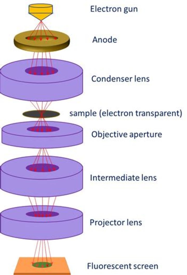
Modern instruments employ two projector lenses (one called the intermediate lens) to permit a greater range of magnification and to provide a greater overall magnification without a commensurate increase in the physical length of the column of the microscope.
What is an electron lens?
A lens that performs action of convergence on electron beams similar to the lens action of an optical lens on light rays. The “electron lens” is classified into the magnetic-field type and the electric-field type. The former is called the electromagnetic lens, and the latter is the electrostatic lens.
Does an electron microscope use a glass lens?
Electron microscope description However, electrons travel as much smaller wavelengths than visible light, enabling them to reveal smaller details than what is possible with light. Instead of a glass condenser lens, electron microscopes use electromagnetic or electrostatic lenses.Why electromagnetic lens is used in electron microscope?
In a similar way to optical microscopes, lenses are used to control the path of the electrons. Because electrons cannot pass through glass, the lenses that are used are electromagnetic.
What is a scanning electron microscope used for?
Because of its great depth of focus, a scanning electron microscope is the EM analog of a stereo light microscope. It provides detailed images of the surfaces of cells and whole organisms that are not possible by TEM. It can also be used for particle counting and size determination, and for process control.
Does an electron microscope use light?
The electron microscope uses a beam of electrons and their wave-like characteristics to magnify an object’s image, unlike the optical microscope that uses visible light to magnify images. … This stream is confined and focused using metal apertures and magnetic lenses into a thin, focused, monochromatic beam.
How do electrostatic lenses work?
An electrostatic lens is a device that assists in the transport of charged particles. For instance, it can guide electrons emitted from a sample to an electron analyzer, analogous to the way an optical lens assists in the transport of light in an optical instrument.How is the image formed in an electron microscope?
An image is formed from the interaction of the electrons with the sample as the beam is transmitted through the specimen. The image is then magnified and focused onto an imaging device, such as a fluorescent screen, a layer of photographic film, or a sensor such as a scintillator attached to a charge-coupled device.
What is electrostatic lens in CRO?Electrostatic lens consists of three anodes, with the middle anode at a lower potential than the other two electrodes. … A pd is kept between these two electrodes so that an electric field is generated between them. Spreading of electric field is caused because of repulsion between electric lines.
Article first time published onHow do electron optics work?
electron optics, branch of physics that is concerned with beams of electrons, their deflection and focusing by electric and magnetic fields, their interference when crossing each other, and their diffraction or bending when passing very near matter or through the spacings in its submicroscopic structure.
What is a condenser lens on a microscope?
On upright microscopes, the condenser is located beneath the stage and serves to gather wavefronts from the microscope light source and concentrate them into a cone of light that illuminates the specimen with uniform intensity over the entire viewfield.
What type of microscope uses magnetic lenses to bend beams of electrons?
This device is called a magnetic lens because it bends the beam of electrons the same way that a glass lens bends a beam of light. A scanning electron microscope usually has more than one lens.
Can we see atoms with an electron microscope?
“So we can regularly see single atoms and atomic columns.” That’s because electron microscopes use a beam of electrons rather than photons, as you’d find in a regular light microscope. As electrons have a much shorter wavelength than photons, you can get much greater magnification and better resolution.
Why can electron microscope detect more detail?
Electron microscopes differ from light microscopes in that they produce an image of a specimen by using a beam of electrons rather than a beam of light. Electrons have much a shorter wavelength than visible light, and this allows electron microscopes to produce higher-resolution images than standard light microscopes.
What type of image does a scanning electron microscope produce?
A scanning electron microscope (SEM) is a type of microscope which uses a focused beam of electrons to scan a surface of a sample to create a high resolution image. SEM produces images that can show information on a material’s surface composition and topography.
Does scanning electron microscope produce 3D images?
SEMs do not naturally provide 3D images contrary to SPMs. However 3D data can be obtained using an SEM with different methods as follows.
What kind of light is used in optical microscope?
The optical microscope, also referred to as a light microscope, is a type of microscope that commonly uses visible light and a system of lenses to generate magnified images of small objects.
What is diffraction contrast?
Diffraction contrast means the intensity change in an electron microscope image that is formed when the diffraction condition is changed with areas of the specimen. In the bright-field image (formed by the transmitted wave), the area where diffraction takes place loses its image intensity, thus getting dark.
What is the resolution of electron microscope?
The wavelength of electrons is much smaller than that of photons (2.5 pm at 200 keV). Thus the resolution of an electron microscope is theoretically unlimited for imaging cellular structure or proteins. Practically, the resolution is limited to ~0.1 nm due to the objective lens system in electron microscopes.
Which type of electrons are used in image formation of tem?
TEMs employ a high voltage electron beam in order to create an image. An electron gun at the top of a TEM emits electrons that travel through the microscope’s vacuum tube.
What is ion lens?
ION Lens is a companion application that acts as an interface or a display unit for the battery pack through a battery management system. … It provides the end-user of a stationary storage system or an EV with complete control and convenience!
What are ion optics?
the branch of science that studies the behavior of electron and ion beams in a vacuum under the influence of electric and magnetic fields. Such images are commonly called electron-optical and ion-optical images. …
What is focusing anode in CRT?
The focusing anode is located between pre-accelerating and accelerating anode. The electron while passing through the control grid is accelerated by a high positive potential which is applied to the pre-accelerating or accelerating nodes.
What is an electron lens compare between electron lens and optical lens?
Differences between Light Microscope and Electron MicroscopeLight MicroscopeElectron MicroscopeLive or Dead specimen may be seen.Only Dead or Dried specimens are seen.Condenser, Objective and eye piece lenses are made up of glasses.All lenses are electromagnetic.
Who invented electron microscope?
The invention of the electron microscope by Max Knoll and Ernst Ruska at the Berlin Technische Hochschule in 1931 finally overcame the barrier to higher resolution that had been imposed by the limitations of visible light. Since then resolution has defined the progress of the technology.
Why condenser is used in microscope?
Condensers are located above the light source and under the sample in an upright microscope, and above the stage and below the light source in an inverted microscope. They act to gather light from the microscope’s light source and concentrate it into a cone of light that illuminates the specimen.
What is objective lens in microscope?
The objective lens consists of several lenses to magnify an object and project a larger image. According to the difference of the focal distance, lenses of different magnifications are available, such as 4x, 10x, 40x, and 50x.
What is collector lens?
The base of the microscope contains a COLLECTOR LENS. This lens is placed in front of the light source. Its function is to project an image of the light source onto the plane of the condenser’s aperture dia- phragm.
Do atoms have Colour?
atoms (as opposed to molecules) do not have colors – they are clear except under special conditions..
Do electrons orbit the nucleus?
Unlike planets orbiting the Sun, electrons cannot be at any arbitrary distance from the nucleus; they can exist only in certain specific locations called allowed orbits. … The electron travels in circular orbits around the nucleus. The orbits have quantized sizes and energies.